Researchers at the University of Oxford have unveiled a pioneering method that has the potential to offer customized solutions for individuals affected by brain injuries.

(Photo : FRED TANNEAU/AFP via Getty Images)
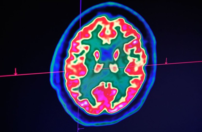
A picture of a human brain taken by a positron emission tomography scanner, also called PET scan, is seen on a screen on January 9, 2019, at the Regional and University Hospital Center of Brest (CRHU - Centre Hospitalier Régional et Universitaire de Brest), western France.
Emerging 3D Printing in Biotechnology
In a groundbreaking achievement, the scientists showcased the ability to 3D print neural cells, replicating the intricate structure of the cerebral cortex for the very first time, as reported by Interesting Engineering.
The emerging domain of 3D printing in biotechnology involves the meticulous construction of living tissue frameworks, employing specialized 3D printers that layer materials systematically.
This innovative technology carries substantial promise across diverse sectors, with particular significance in the realms of medicine and scientific exploration.
Prior to recent advancements, a significant challenge existed: ensuring that 3D-printed stem cells could accurately replicate the intricate architecture of the human brain, leaving a critical gap in this field of research.
Dr. Yongcheng Jin, the lead author affiliated with the Department of Chemistry at the University of Oxford, emphasized the groundbreaking nature of this advancement, which represents a major step forward in creating materials that closely replicate both the structure and function of natural brain tissues.
This development not only opens up a unique avenue for in-depth exploration of the human cortex but also offers a promising prospect for individuals who have suffered brain injuries in the long term.
The cortical structure was meticulously constructed using human induced pluripotent stem cells (hiPSCs), known for their remarkable capability to generate various cell types found throughout the human body.
Employing hiPSCs
EurekAlert! reported that a significant advantage of employing hiPSCs for tissue repair lies in their easy derivation from a patient's own cells, mitigating concerns related to immune responses.
The process involved differentiating hiPSCs into neural progenitor cells to create two distinct layers resembling the cerebral cortex. This transformation was achieved through the precise application of specific growth factors and chemical combinations.
The resulting cells were then suspended in a solution, resulting in the creation of two distinct "bioinks" that served as the basis for the subsequent 3D printing process, ultimately yielding a two-layered tissue structure.
In laboratory settings, these printed tissues exhibited remarkable resilience, maintaining their layered cellular arrangement for extended periods. This enduring structural integrity was affirmed through the consistent expression of layer-specific biomarkers.
Upon implantation into mouse brain slices, the printed tissues exhibited impressive integration capabilities. This was evidenced by the projection of neural processes and the migration of neurons across the interface between the implant and the host tissue.
Notably, the implanted cells displayed signaling activity, indicating effective communication between human and mouse cells and thereby demonstrating both functional and structural integration.
Looking forward, the research team aims to further enhance the droplet printing technique to create more intricate, multi-layered cerebral cortex tissues that more accurately replicate the architectural complexity of the human brain.
Beyond their potential applications in repairing brain injuries, these engineered tissues hold promise for drug evaluation, research into brain development, and enhancing our understanding of the foundations of cognition. The findings of the study has been published in the journal Nature Communications.
Related Article : How the Human Brain Is Inspiring AI Systems to Be More Robust


