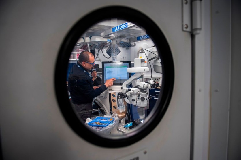
Researchers have made significant strides in understanding the eye's molecular age and health by examining eye fluid. The study aimed to bridge the gap between visible eye changes and molecular alterations in eye fluid, employing non-invasive liquid biopsy methods to collect vital samples without harming the eye.

Unlocking Secrets of Eye Health and Aging
In a recent investigation, scientists have unveiled intriguing insights about the eye's molecular age and health status by scrutinizing the composition of eye fluid.
The study involved the analysis of minuscule liquid droplets, typically discarded during eye surgeries, where nearly 6,000 proteins originating from diverse cell types within the eye were discovered.
The study's lead author, Vinit Mahajan, highlighted the eye's unique role as an organ that provides real-time insights into the development of diseases.
Mahajan noted that the study aims to bridge the observable changes in the eye with the molecular alterations detected in the eye fluid. The research team encountered a formidable challenge when attempting to sample the minute eye fluid.
Traditional tissue sampling methods were not a viable option due to the potential harm they could inflict on the eye. Instead, they turned to liquid biopsy techniques, which involve the collection of fluid from the vicinity of cells or tissues of interest.
However, Interesting Engineering reported that prior liquid biopsy methods faced limitations in quantifying numerous proteins and pinpointing their cellular origins, which are pivotal for diagnosis and treatment.
To overcome this hurdle, Mahajan's team group employed a high-resolution methodology, which enabled them to analyze 120 liquid biopsies sourced from the aqueous or vitreous humor of patients who underwent eye surgery. Remarkably, they identified 5,953 proteins - 10 times more than previous studies.
Proteomic Clock
The research team then harnessed the capabilities of an AI-driven machine learning model to construct what they've termed a "proteomic clock."
This innovative clock is designed to estimate the molecular age of the eye, relying on a dataset of 26 essential proteins. The model demonstrated remarkable accuracy when applied to healthy eyes.
However, it unveiled a remarkable insight - conditions such as diabetic retinopathy and uveitis could induce accelerated aging in specific cell types within the eye.
To illustrate, individuals afflicted with severe diabetic retinopathy displayed eyes that appeared to be 30 years older than their actual chronological age.
Intriguingly, EurekAlert reported that these signs of accelerated aging often manifested even before the onset of disease symptoms and continued to persist following treatment. The research study unearthed the presence of proteins in the eye fluid that are typically only detectable after death.
These proteins are vital for diagnosing Parkinson's disease, a condition notorious for its diagnostic challenges. Present diagnostic tests lack the capability to measure these proteins, making early diagnosis elusive.
The study's authors have drawn several noteworthy conclusions, Cell reported. They underscored that the aging process is far from uniform across different organs and cell types. This finding bears significant implications for the realm of precision medicine and the design of clinical trials.
The researchers outlined their future research trajectory. Their next endeavor involves the comprehensive characterization of samples obtained from a broader spectrum of patients and encompassing various eye-related diseases.
Additionally, the researchers envision the potential application of their methodology to other tissues that pose challenges for traditional sampling techniques.


