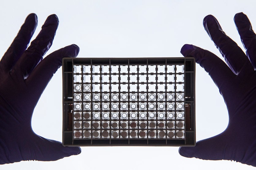
A new development emerges as a silicon-based biochip enables rapid genetic screening of numerous molecules, with the potential to identify over 160,000 distinct entities in a single square centimeter.

Enabling Swift Detection to Cancer Protein Markers, Viruses
A groundbreaking achievement emerges from the scientific community as a minute biochip, constructed from silicon blocks, showcases the capability to perform swift genetic screening on a multitude of molecules. As reported in Science, this tool holds the capacity to potentially discern more than 160,000 unique molecules within a single square centimeter.
The far-reaching implications of this innovation span across various medical domains. Notably, it offers avenues for detecting cancer protein markers and facilitating clinical diagnoses of respiratory infections. This pioneering technology introduces a novel dimension to medical advancement, providing a versatile tool to address critical challenges in healthcare.
A cornerstone of many genetic test sensors involves observing the absorption or emission of light from specific molecules engineered to attach to the desired gene. This technique commonly relies on the polymerase chain reaction to replicate a multitude of copies of the target prior to identification, thereby contributing to heightened expenses and an extended testing timeline.
Furthermore, earlier iterations of genetic screening sensors were limited in their ability to discern diverse target compounds and necessitated the use of optical markers for target sequence detection.
Developing the Tool
The study authored by researchers from Stanford University introduces a genetic screening platform that operates without the need for labeling. Interesting Engineering reported that this innovative approach harnesses high-quality (high-Q) factor silicon nanoantennas that have been modified with nucleic acid fragments.
In developing this tool, the researchers harnessed an optical detection technology centered around metasurfaces constructed from diminutive silicon boxes. These minuscule silicon arrays possess dimensions of roughly 500 nanometers in height, 600 nanometers in length, and 160 nanometers in width.
The top surface of the silicon boxes has the capacity to concentrate near-infrared light, courtesy of the nanoantennas. Elaborating on this phenomenon, the study explains that these metasurfaces incorporate subwavelength nanoantennas, adept at confining light in the vicinity while simultaneously offering meticulous command over the scattering of light in the distant field.
Amidst the steady progression of advancements in cancer detection, researchers hailing from the Massachusetts Institute of Technology (MIT) have unveiled an innovative wearable ultrasound device, poised to redefine the landscape of breast cancer detection.
With the potential to usher in early diagnoses and enhanced survival rates, this development stands as a beacon of hope. The disparity in survival rates between early-stage and late-stage breast cancer diagnoses underscores the criticality of timely detection. Recognizing this imperative, the researchers have ingeniously engineered a pliable patch that seamlessly affixes to undergarments.
This wearable patch empowers individuals to conduct ultrasound imaging on their breast tissue, affording the flexibility to explore various imaging angles. This innovation holds the promise of transforming how breast cancer is detected, offering a proactive approach to diagnosis that could potentially save countless lives.
Related Article : New Wearable Ultrasound Scanner That Fits in Bra Can Help Detect Breast Cancer Early

![Apple Watch Series 10 [GPS 42mm]](https://d.techtimes.com/en/full/453899/apple-watch-series-10-gps-42mm.jpg?w=184&h=103&f=9fb3c2ea2db928c663d1d2eadbcb3e52)


