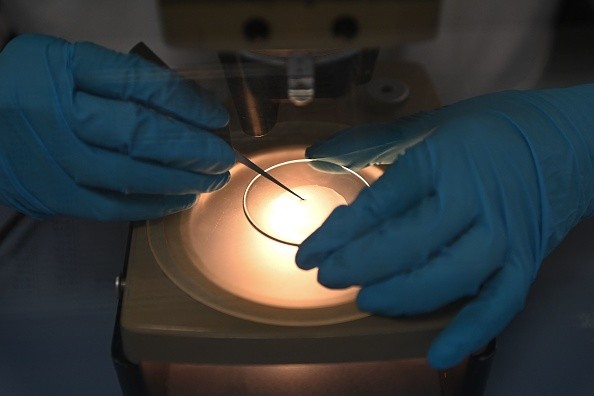
Researchers from South Korea have developed a new holographic microscope that provides 3D imaging and high resolution of a living mouse's brain, Interesting Engineering reported.

On July 28, this research was published, and the development was led by two researchers, Associate Director of the Center for Molecular Spectroscopy and Dynamics within the Institute for Basic Science Choi Wonshik and The Catholic University of Seoul National University Professor Kim Moonseok.
The demonstration of the new invention was used in observing a living mouse's brain. Science Daily reported that the observation succeeded as the researchers obtained an image that was clear with high-resolution quality.
Sharp images, especially high-resolution results, were very hard to obtain in usually used microscopes as multiple scattering effects and severe aberration for living issues will only occur when a certain light hits the cells.
Assessing the signal reflected from the target tissue and putting enough light energy into the sample is very important because the interior properties of living organisms will be explored using light.
How Did It Improve?
Future scientists will be able to experience a much better experience in the newly developed microscope as the wavefront distortion of the light from the object that they are examining is already corrected. This is also possible even if only a minimal light was used during the procedure.
A statement from Institute for Basic Science (IBS) was released and stated that this process would have various scattering effects. Because of this, reducing the ratio of multiple-scattered waves and raising the resolution of the deep tissue will be hard to obtain despite the development.
Also Read : Quantum Microscope to Soon Help Scientists See 'Impossible' Objects Under the Instrument in New Study
Researchers have also made a method for the selection of single-scattered waves as they have similar reflection waveforms no matter what angle will be positioned as per the results that can be obtained online on Science Advances.
This will focus on more than 80 times the light energy on the neural fibers from past inventions, as they used complex algorithms and an operation that analyzes the eigenmode of a medium. Adding to this is the microscope will selectively remove the unnecessary signal and will only focus on the important visuals.
As per Professor Moonseok and Doctor Yongheyon, "When we first observed the optical resonance of complex media, our work received great attention from academia. From basic principles to practical application of observing the neural network beneath the mouse skull, we have opened a new way for brain neuroimaging convergent technology by combining the efforts of talented people in physics, life, and brain science."
Related Article : Researchers Developed Nanopipes That Could Help in Better Drug Delivery to the Human Body
This article is owned by TechTimes
Written by Inno Flores
![Apple Watch Series 10 [GPS 42mm]](https://d.techtimes.com/en/full/453899/apple-watch-series-10-gps-42mm.jpg?w=184&h=103&f=9fb3c2ea2db928c663d1d2eadbcb3e52)


