Despite advances in cardiovascular disease research in the past decades, heart disease remains the leading cause of death worldwide. In fact, sudden cardiac death (SCD) alone accounts for up to 20% of all fatalities.
But an ethical, more precise alternative to current methods might be provided by a miniature working model of a human ventricle. This new model could pave the way for the creation of novel medications and treatments and further the study of cardiovascular conditions, according to a report by ScienceAlert.

A Miniature Exit Chamber of a Heart
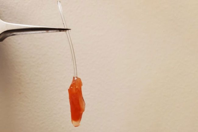
A millimeter-long (0.04 inches) vessel that replicates the muscular exit chamber of a human embryo's heart was reverse-engineered by scientists from the University of Toronto and the University of Montreal in Canada. It not only beats realistically but also pumps fluid!
The left ventricle of the human heart is responsible for delivering newly oxygenated blood to the aorta and then to all the parts of the body.
Sargol Okhovatian, a biomedical engineer at the University of Toronto, said that the new model can monitor both the fluid pressure and ejection volume, which how much fluid is pushed out each time the ventricle contracts.
It is worth noting that there are often only a few alternatives for researching how a healthy or sick heart distributes blood.
ScienceAlert stated that authentic models can be provided by non-functional organs, such as those that were removed during an autopsy. Although tissue cultures may offer a glimpse into biochemical operation, they fall short of replicating the hydraulics of a three-dimensional, pulsing mass.
Although it's not necessarily the most ethical decision, using an animal model also enables scientists to evaluate how a beating heart works as a pump when affected by novel treatments. But this new model could expand alternatives.
How The Heart Was Made
This new heart-like organ was produced in a lab using a combination of synthetic and biological materials, young rats' cardiovascular tissues served as the source of the cells, which were subsequently grown on a layer of scaffold printed from a polymer with grooves to guide the growth of the tissue.
The massive final chamber that squeezes blood into the aorta with one powerful blow was pushed to resemble the alignment of heart muscle fibers by the new model.
The team utilized a cone-shaped shaft they called a mandrel to transform the triple-layered stack of heart cells into a model that mimics a pulsing chamber.
The vessel's interior diameter of less than half a millimeter (0.02 inches) and pressure of about 5% of an adult's heart hardly allow it to expel fluids but the model is still a proof of concept that might eventually be expanded to include more tissue layers and depict a more robust system.
Without the need for invasive surgery or animal testing, these models could pave the way to examine not only cell function but also tissue and organ function, according to senior author Milica Radisic, a chemist from the University of Toronto.
This article is owned by Tech Times
Written by Joaquin Victor Tacla
ⓒ 2025 TECHTIMES.com All rights reserved. Do not reproduce without permission.


